Extensive Amalgam Tattoo (Amalgam Pigmentation) on the Palatal Mucosa: A Case Report
Abstract
Introduction: Amalgam tattoo is the most common exogenous oral pigmentation, caused by traumatic implantation of dental amalgam into soft tissue.
Observation: We report a case of large amalgam pigmentation on the right hard palate.
Discussion: Amalgam tattoo can sometimes be confused with melanotic lesions, being then biopsied. Once the diagnosis of amalgam tattoos has been established, the removal of lesions is not necessary, except for esthetic reasons.
Keywords: Amalgam tattoo; Oral mucosa; Pigmentation.
Introduction
Oral pigmentations may be classified into two major groups on the basis of their clinical appearance: focal and diffuse pigmentations. All pigmented oral cavity lesions should be viewed with suspicion to eliminate a malignant melanoma. This article deals with an extensive amalgam tattoo lesion on palatal mucosa which required a biopsy for a definitive diagnosis.
Case Presentation
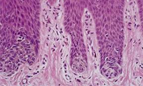
A 56-year-old man with an unremarkable medical history was referred to the department of maxillofacial surgery on suspicion of mucosal melanoma. Clinical examination found a large brown flat macula located on the right hard palate adjacent to a restored tooth 16 with the presence of amalgam fillings (Figure 1). There was no lymphadenopathy. Panoramic X-ray did not show any abnormalities. A biopsy was performed under local anesthesia. Histology showed brownish-black pigment along the collagenous fibers and in the vascular sheaths. No melanocytes or naevus cells were found (Figure 2). The findings suggested the diagnosis of amalgam tattoo. The patient didn’t need any further treatment.
Discussion
Amalgam tattoos are common exogenous pigmented lesions of the oral mucosa occurring mainly by accidental displacement of metal particles in oral soft tissues during restorative dental procedures using amalgam. The diagnosis is simple, clinically amalgam tattoo presents as a slate-gray, bluish or black maculae, Lesions are usually well circumscribed, uniformly pigmented, measuring from 0.1 to 2 cm [1], they affect mainly the mandibular gingival mucosa, followed by the buccal mucosa, floor of mouth, tongue, retromolar mandibular area, lips, and palate [2]. Periapical X-rays may be useful to detect the radio-opacity related to amalgam. However, less than 25% of amalgam pigmentations are radio-opaque since their metallic particles are very small or are too dispersed to be visible in a radiographic study [2,3].


Comments
Post a Comment